For centuries now, humankind has tried to understand the cell. The advent of microscopy has provided a huge boost to what we know about the cell – how it looks and how it works. As technology progressed, microscopy also evolved. Nowadays, we combine advanced imaging techniques with microscopy to develop better images of cells and understand cellular structures in more detail. A recent advancement in the field of cellular analysis is the development of live cell imaging.
What is live cell imaging?
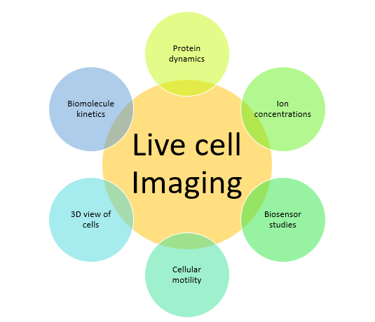
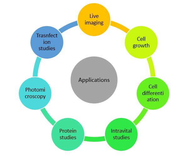
Earlier, cellular imaging involved visualizing fixed cell specimens under microscopes. But the process of fixation kills the cells and not much about the functions of the living cells can be observed. For example, what if we wanted to study the motility of the cell? Or maybe the movement of cellular structures within a live cell? Live cell imaging is a solution to these problems. Since this technique does not kill the cells, it allows one to track dynamic changes within the cell over a period of time. Some of the applications of live cell imaging are shown in the image below:
For growing cells under in-vitro conditions in laboratory, cell cultures are maintained inside incubators. Incubators are special devices which are used to provide the right environment for the growth of cells, in terms of temperature, humidity and carbon dioxide concentration. It would be most ideal if one could image the cells in a culture when they are inside the incubator as it would allow one to study the changes in a cell over hours or even days. This thought is what gave rise to live cell imaging inside incubators.
Currently, several technologies are available for live cell imaging within incubators. Three main strategies are followed during live cell analysis – automated monitoring and control of cell culture conditions, automated microscopy, and combination of the two.
Currently, several companies provide solutions for live cell imaging in life sciences and pharmaceutical laboratories. Let’s discuss how a few of them work.
The CytoSMART Lux2 and Omni system
As we discussed earlier, living cells are dynamic systems and studying them in real-time would be much more beneficial than end-point analysis. The CytoSMART systems are designed to allow a user to observe cell cultures from inside an incubator for extended periods of time – from hours to weeks.
The CytoSMART Omni system
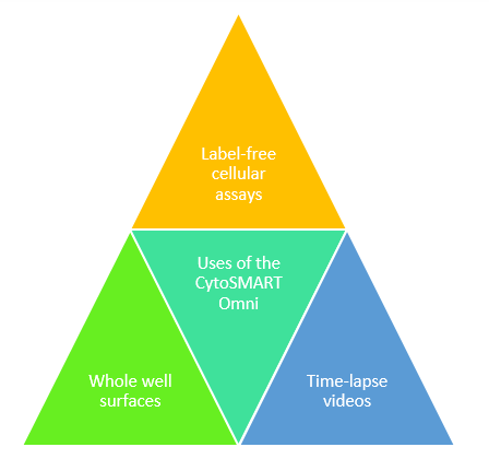
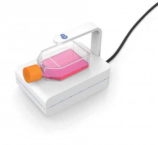
The CytoSMART Omni system is an imaging system that fits inside a cell culture incubator. The system can be used for the analysis of various cell and tissue culture vessels like plates, plates with wells, petri dishes, etc. The system is equipped with an automated bright-field microscope system, with a 10X fixed objective, which can create time-lapse videos for the analysis of changes occurring within a cell over a passage of time. With the help of the Omni system, scientists can observe cell proliferation, wound healing and colony formation assays with greater precision and also glean more information from the assays, than what could be understood from end-point analysis. The system exports data in the JPG, XLSX and MP4 formats, which are compatible with most image analysis software.
The CytoSMART Lux2 system
This system is similar to the CytoSMART Omni, but is smaller. The CytoSMART Lux2 system is designed to allow the overall analysis of the characteristics of the cell culture system. This system also generates time-lapse videos with the help of a 10X objective, bright-field microscope equipped with digital phase-contrast. The Lux2 system is compatible with petri dishes, T-25 flasks and 6-96 well plates. The system is mainly designed to study the health and growth rate of a cell culture, which can provide valuable information regarding sub-culturing time, cellular morphology, cytotoxicity and proliferation rates.
Etaluma’s Lumascopes
Etaluma has developed a live cell imaging system called the Lumascope. This unique system fits inside the standard incubator and can be used for imaging cultures kept in microplates, flasks and other labware. Etaluma has developed three systems – the manual Lumascope 560, Lumascope 620 and the automated Lumascope 720. The system has a phase contrast, an inverted, fluorescent microscope equipped with brightfield and fluorescence filters of different colours. The microscope also has a variable objective. The imaging system uses advanced optical systems, CMOS sensors, LED light-sources, and filters to give high quality images. The microscope has an XY stage, in case of the manual systems and the automated systems have an XYZ stage. Etaluma also provides a proprietary software called the Lumaview that facilitates image analysis. The following image summarizes the uses of the Lumascope system:
Biotek’s Live Cell Imaging System
Biotek combines two technologies – the BioSpaTM8 and the CytationTM5 Cell Imaging Multi-Mode Reader, to provide live cell imaging solutions for cellular and molecular studies. The BioSpaTM8 is essentially an automated incubator in which cell cultures can be stored and efficiently monitored. The system is compatible with cell culture flasks, petri plates and other labware.
The CytationTM5 is an automated image capturing system. The microscope has an adjustable objective of up to 60X and the imaging system includes fluorescence filters, phase contrast and brightfield microscopy. The system is equipped with a robotic arm that transfers cell culture vessels from the BioSpaTM8 to the CytationTM5 Cell Imager. The analysis system contains several options that allow time-lapse generation, Z-stacking, projections, phase contrast, image stitching, etc.
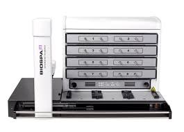
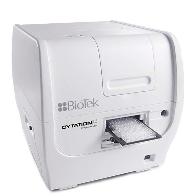
The BioSpaTM8 and the CytationTM5 Cell Imaging Multi-Mode Reader (https://www.news-medical.net/)
Celldiscoverer7
The Cellsdiscoverer7 is an automated microscope by Ziess. This boxed microscope system works like an inverted microscope. Zeiss also provides an incubator that can be used for cell culture. The incubator provides the required cell culture conditions and it minimizes the manual adjustment of = conditions. The Celldiscoverer7 microscope has a variable objective. This system is compatible with the Airyscan 2 which allows confocal and widefield imaging. The dynamic process can be easily analysed using this system using FRAP and FRET, etc.
Live-cell imaging is a new era in microscopy and is turning to be great boost to increasing our knowledge of cellular systems. This technique is now regularly used in life sciences, pharmaceutical industry and research. Especially in fields like cancer biology, it is helping scientists get a deeper understanding of the cellular system, about how cancer cells are different from normal cells. Live-cell imaging can go a long way in improving research methodologies and outcomes.
References:
- https://www.labome.com/method/Live-Cell-Imaging.html
- https://www.cytosmart.com/products/omni
- https://cytosmart.com/products/lux2
- https://etaluma.com/products/about-lumascope-fluorescent-microscopes/
- https://www.zeiss.com/microscopy/int/products/imaging-systems/celldiscoverer-7-automated-microscope-for-live-cell-imaging.html
- https://www.biotek.com/products/imaging-microscopy-cell-imaging-multi-mode-readers/cytation-5-cell-imaging-multi-mode-reader/